
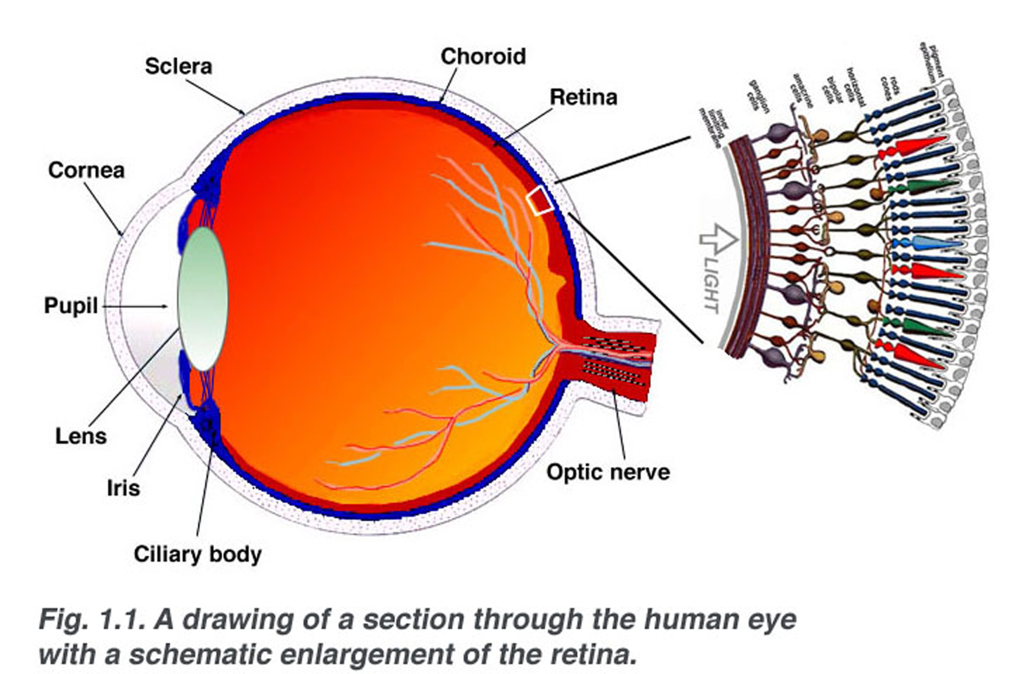
The retina is a thin layer of tissue that lines the back
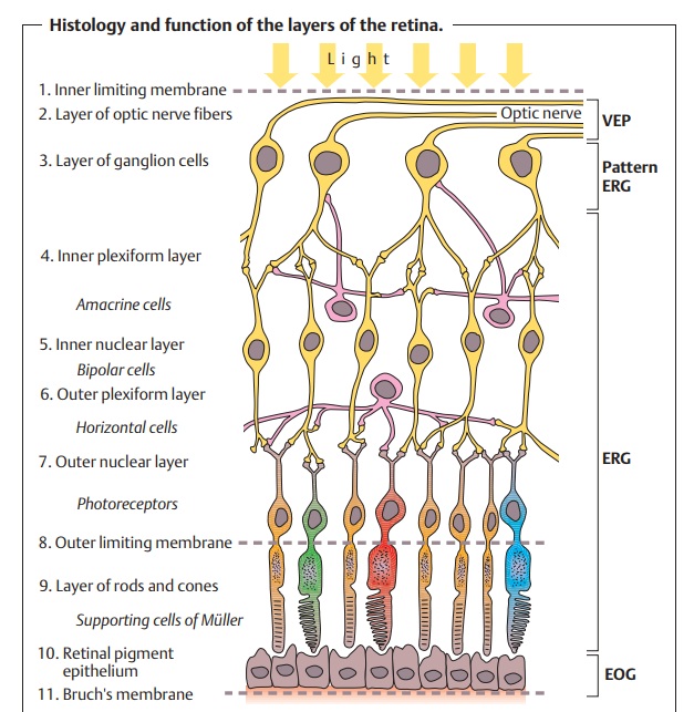
Ten layers of cells in the retina can be seen microscopically. In general, there are four main layers: (1) Next to the choroid is the pigment epithelium, already mentioned. (2) Above the epithelium is the layer of rods and cones, the light-sensitive cells.

Gross Anatomy Of Retina ANATOMY
The retina consists of layers, which can be subcategorised into retinal pigmented epithelium (RPE) and neural retina. The RPE is a single layer of cuboidal epithelial cells and located in the outermost layer of the retina. It is responsible for the nourishment and support of the neural retina. The tight junctions between the RPE cells form part.

The basic retinal structure. Histological appearance of choroid and... Download Scientific Diagram
The retina is the layer of cells lining the back wall inside the eye. This layer senses light and sends signals to the brain so you can see. Several parts of the eye are associated with the retina. They include: Peripheral retina Macula Fovea Photoreceptors Rods Cones Eye problems that can affect the retina include: Branch retinal vein occlusion
Simple Anatomy of the Retina by Helga Kolb Webvision
Retinal pigment epithelium - This is a single layer of cells that provide essential nutrition and waste removal for the photoreceptor cells. Accumulation of waste can lead to AMD and Stargardt disease. Photorecptors - This is where the rods and cones are located that convert light into electrical signals.

A schematic of the retina showing overall arrangement of retinal layers...
The retina is a layer of photoreceptors cells and glial cells within the eye that captures incoming photons and transmits them along neuronal pathways as both electrical and chemical signals for the brain to perceive a visual picture.

Human eye Retina, Optic Nerve, Vision Britannica
The arteries pierce the sclera around the optic nerve and fan out to form the three vascular layers in the choroid: outer (most scleral), medial and inner (nearest Bruch's membrane of the pigment epithelium) layers of blood vessels. This is clearly shown in the corrosion cast of a cut face of the human choroid in ).

Retinal layers. Retina is formed by 10 layers from the inner to the... Download Scientific Diagram
The layers 1-6 of retina are supplied by the branches of the central retinal artery, while the layers 7-10 are supplied by the capillaries from the choroid. Refractive media of the eyeball The refractive media of the eye are the structures that help in focusing the ray of light onto the retina where it can be detected by the photoreceptors. The.

How the Retina Works Detailed Illustration Eye health, Eye anatomy, The retina
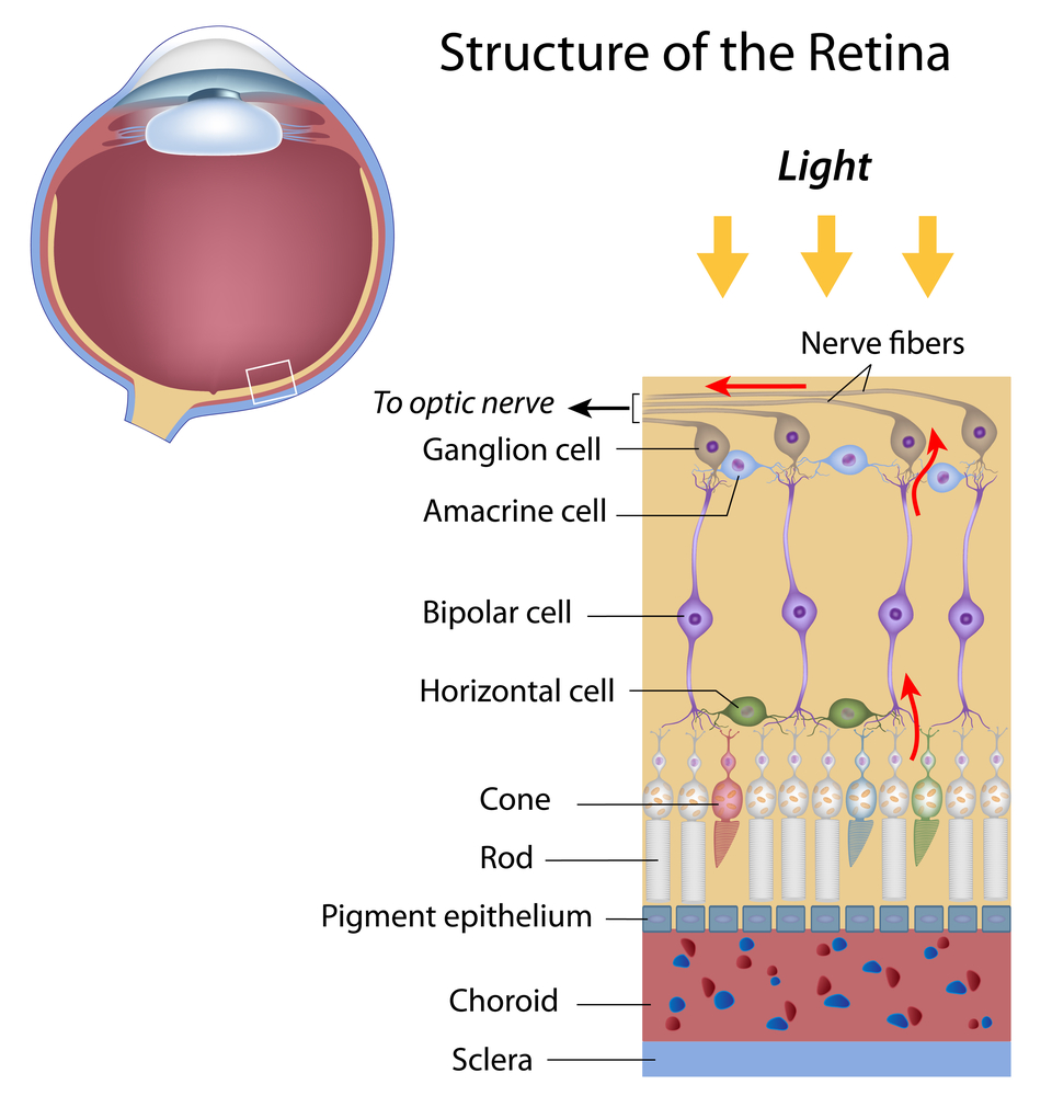
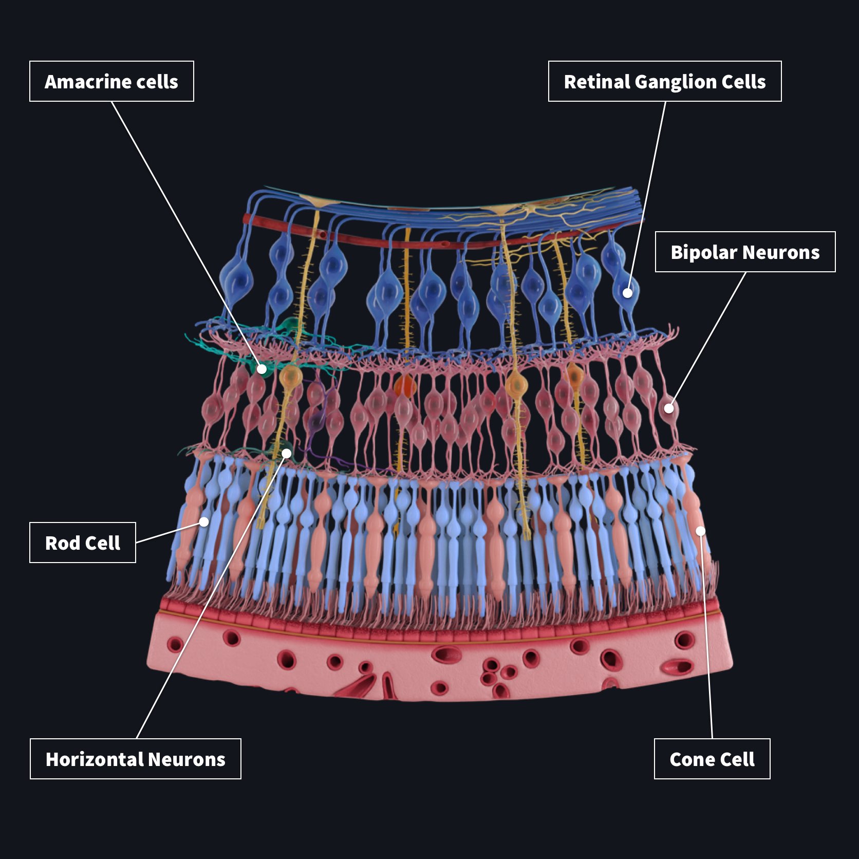
The neural retina consists of several layers of neurons interconnected by synapses and is supported by an outer layer of pigmented epithelial cells. The primary light-sensing cells in the retina are the photoreceptor cells, which are of two types: rods and cones. Rods function mainly in dim light and provide monochromatic vision.

Layers of the Retina Download Scientific Diagram
Tears lubricate the eye and are made up of three layers. These three layers together are called the tear film. The mucous layer is made by the conjunctiva. The watery part of the tears is made by the lacrimal gland. The eye's lacrimal gland sits under the outside edge of the eyebrow (away from the nose) in the orbit.

Retinal epithelial layers The retina, Eye anatomy, Eye health
The retina is a thin layer of tissue that lines the very back of the inside of the eyeball. The retina contains millions of cells that perceive light, color, and fine details in the things you see. A number of diseases can affect the retina, including cancer. If any part of the retina becomes damaged, your vision may be compromised.
Layers of the Retina Discovery Eye Foundation
The pigment epithelium is the most external layer of the retina. It abuts on the choroidal layer of the eye. It contains a single layer of cuboidal-supporting cells for the neural portion of the retina. These cells contain melanin, which absorbs light and decreases light scatter within the eye.

Schematic crosssection showing the retinal blood vessels lining the... Download Scientific
The retina is composed of epithelial, glial, and neural cells that are organized into 10 distinctive layers. Out of these, the first 9 layers belong to the inner neurosensory retina, one of which are the photoreceptors that are sensitive to light.
SNEAK PREVIEW Retinal Layers Complete Anatomy
Discover a world of convenience with our Clearance. Best sellers up to 90% off. Come and check everything at a surprisingly low price, you'd never want to miss it.
/GettyImages-308783-003-56acdcd85f9b58b7d00ac8e8.jpg)
The Anatomy of the Retina
Anatomy of the Retina. The human retina consists of layers of neural tissue that line the entire back wall of the eye. It's the only extension of the brain visible from outside the body (via a retinal exam). 1. The retina attaches to the optic nerve at the optic disc. The optic nerve is one of the main cranial nerves coming from the brain.

Gross Anatomy Of Retina ANATOMY
Labeled anatomy of the retina and eye. What is the retina of the eye? The retina converts light that enters into your eye into electrical signals your optic nerve sends to your brain which creates the images you see. It's a key part of your vision. The retina is the layer at the very back of your eyeball. Advertisement
Retina
The ten layers of the retina from interior (bordering vitreous humor) to exterior (bordering choroid and sclera) are listed and described below.4,5 Inner Limiting Membrane - forms a barrier between the vitreous humor and the neurosensory retina.